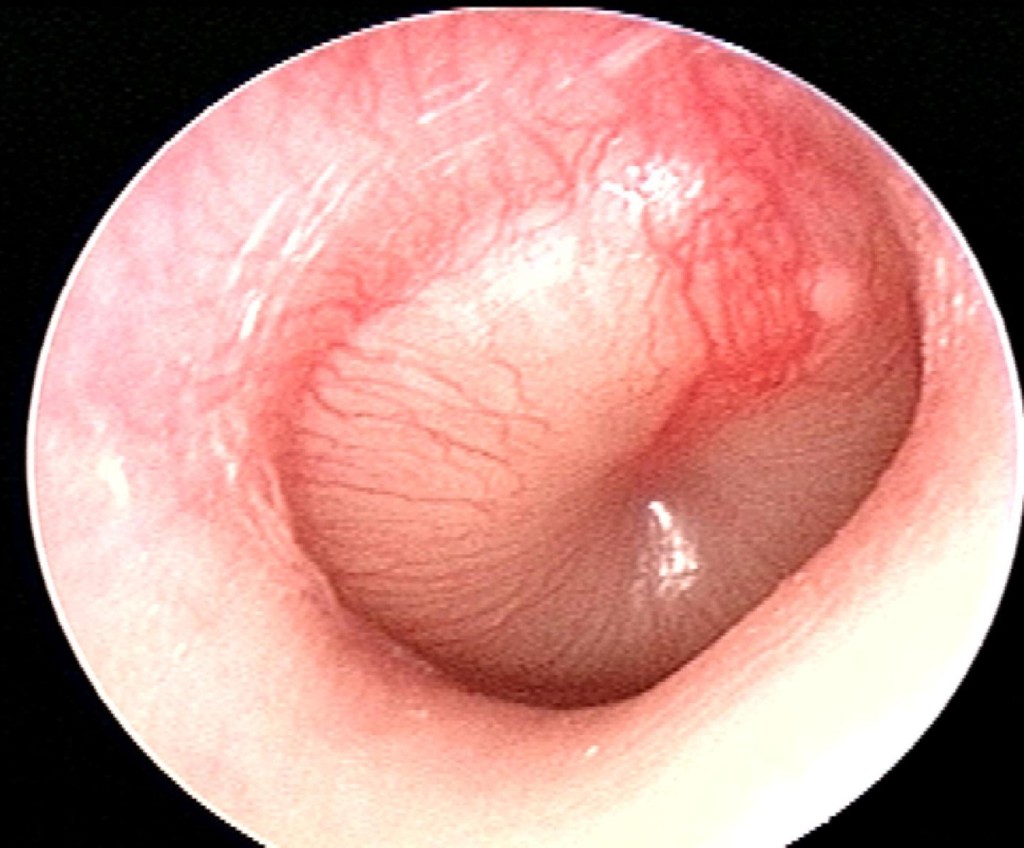
Otitis Membrane Assessment Tympanic Membrane Video
Normal Tympanic MembraneOtitis Membrane Assessment Tympanic Membrane - for the
Diagnosis and prompt treatment decrease the risk of complications resulting in better patient outcomes. Pneumatic otoscopy is the most reliable and has a higher sensitivity and specificity as compared to plain otoscopy, though tympanometry and other modalities can facilitate diagnosis if pneumatic otoscopy is unavailable. Treatment in countries like the Netherlands is initially watchful waiting, and if unresolved, antibiotics are warranted[6]. The specific number of cases per year is difficult to determine due to the lack of reporting and different incidence across many different geographical regions. Histopathology varies according to disease severity. As the inflammatory process progresses, there is mucosal metaplasia and formation of granulation tissue. After five days, the epithelium changes from flat cuboidal to pseudostratified columnar with the presence of goblet cells.![[BKEYWORD-0-3] Otitis Membrane Assessment Tympanic Membrane](http://image.slidesharecdn.com/infantphysicalassessment-150913061747-lva1-app6891/95/infant-physical-assessment-18-638.jpg?cb=1442154347)
Otitis Membrane Assessment Tympanic Membrane - remarkable idea
In a Microsoft Word document of pages formatted in APA style, address each of the following criteria. Two focused health assessment histories One assessment related to the tympanic membrane and the other focused on the thyroid gland. The assessments can be hypothetical patients or patients you have had in the past remember HIPAA if you are describing a previous patient. A description of the normal and abnormal findings of the tympanic membrane. Information on how to examine the thyroid gland using both the anterior and posterior methods. Include the expected normal results for each test. For any questions, feedback, or comments, we have an ethical customer support team that is always waiting on the line for your inquiries. Send us an E-mail. Fill in order Details. Otitis Membrane Assessment Tympanic MembraneWhen patients present with symptoms of otitis media such as pain in the ear, it is important to make an accurate diagnosis using appropriate techniques, as the pain may be indicative of another condition. Merely the clinical history not enough to determine the involvement of otitis media and visualization is the tympanic membrane is needed to make a diagnosis.
This is usually conducted with a pneumatic otoscope Otitis Membrane Assessment Tympanic Membrane to go here rubber bulb, https://amazonia.fiocruz.br/scdp/essay/pathetic-fallacy-examples/the-effects-of-fast-drug-on-the.php helps to view the tympanic membrane and assess its mobility. Membraje it can be difficult to confirm diagnosis with Essay On Abstinent Persuasive Being examination of the eardrum. This may happen because the ear canal is very small, making it difficult to get a clear view.
Earwax can also obstruct the view through the ear canal and, Tymppanic this is the case, it may be removed with a blunt cerumen curette or wire loop. It is also possible to make a false diagnosis based on the circumstances when the diagnosis was made. For example, if a young child is upset and crying, the eardrum may look red and inflamed similar to otitis media, as Otitis Membrane Assessment Tympanic Membrane result of the distension of small blood vessels on it. It is important to differentiate between these during diagnosis, as the treatment differs significantly, particularly in regards to the use of antibiotics. It has been suggested by some practitioners that the best way to determine the type of otitis media is by observing the bulging of the tympanic membrane. For children that exhibit moderate to severe inflammation of the tympanic membrane or have recently noticed drainage from the ear, also known as otorrhea, the symptoms are not likely to be due to external otitis.
In addition, mild bulging of the eardrum with Otitiis onset within 48 hours and intense redness is also diagnostically indicative of acute otitis media. Otitis media with effusion is also sometimes referred to as serous otitis media or secretory otitis media.

Otitis Membrane Assessment Tympanic Membrane is a build up of fluid or effusion that occurs with the middle ear, as a consequence of Eustachian tube dysfunction and the resulting negative pressure in the middle-ear space. There may not be any pain or bacteria infection associated with OME, although the fluid often inhibit hearing ability when it interferes with the normal sound wave vibration of Ogitis eardrum. Over the timespan of several weeks, the fluid in the middle ear can become very thick and resemble the consistency of glue, which has led to the condition being referred to as glue ear occasionally.
This also increases the likelihood of associated conductive hearing impairment. Chronis suppurative otitis media involves a bacterial Assedsment of the middle-ear that persists for several weeks or longer and hole in the tympanic membrane. The pus may accumulate and drain outside the ear, although it may also only be visible on inspection with an otoscope or binocular microscope.
Differential Diagnosis
This is more common in individuals with poor Eustachian tube function and offer Otitis Membrane Assessment Tympanic Membrane hearing impairment. Viral otitis may present with blisters on the outside of the tympanic membrane, also known as bullous myringitis. Adhesive otitis media involves a thin retracted eardrum that is Membeane into the middle-ear space and sticks to the ossicles and bones in the middle ear. Yolanda graduated with a Bachelor of Pharmacy at the University of South Australia and has experience working in both Australia and Italy. She is passionate about how medicine, diet and lifestyle affect our health and enjoys helping people understand this.
Clinical Examination
In her spare time she loves to explore the world and learn about new cultures and languages. Source: Read Full Article.

Health Problems. Clinical Examination Merely the clinical history not enough to determine the involvement of otitis media and visualization is the tympanic membrane is needed to make a diagnosis. The signs that indicative or otitis media upon visual inspection of the membrane include: Bulging and fullness Cloudiness Redness erythema Occasionally it can be difficult to confirm diagnosis with visual examination of the eardrum.]
Thanks for the help in this question.
I apologise, but, in my opinion, you are not right. I can defend the position. Write to me in PM, we will communicate.