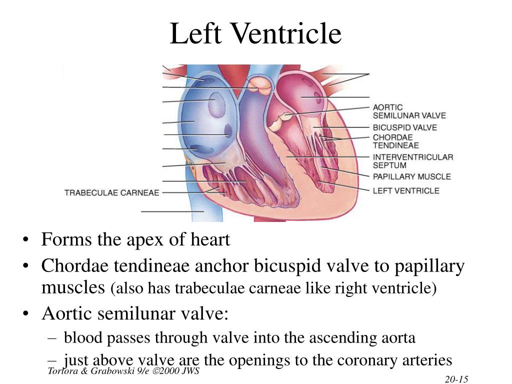
![[BKEYWORD-0-3] The Left Ventricle of the Heart](https://classconnection.s3.amazonaws.com/246/flashcards/543246/png/left_ventricle1321041195695.png)
The Left Ventricle of the Heart - recommend
The measurement of left ventricular ejection fraction LVEF is a ubiquitous component of imaging studies used to evaluate patients with cardiac conditions and acts as an arbiter for many management decisions. This follows early trials investigating heart failure therapies which used a binary LVEF cut-off to select patients with the worst prognosis, who may gain the most benefit. Forty years on, the cardiac disease landscape has changed. Left ventricular ejection fraction is now a poor indicator of prognosis for many heart failure patients; specifically, for the half of patients with heart failure and truly preserved ejection fraction HF-PEF. It is also recognized that LVEF may remain normal amongst patients with valvular heart disease who have significant myocardial dysfunction. This emphasizes the importance of the interaction between LVEF and left ventricular geometry. Guidelines based on LVEF may therefore miss a proportion of patients who would benefit from early intervention to prevent further myocardial decompensation and future adverse outcomes. The assessment of myocardial strain, or intrinsic deformation, holds promise to improve these issues. The measurement of global longitudinal strain GLS has consistently been shown to improve the risk stratification of patients with heart failure and identify patients with valvular heart disease who have myocardial decompensation despite preserved LVEF and an increased risk of adverse outcomes. The Left Ventricle of the Heart.Left ventricular LV dimensions and function can be assessed qualitatively with a handheld echocardiography HHE device. Left ventricular size is reduced when the ventricular cavity appears small in an under-filled patient. On the other hand, LV dimensions may be increased when there is LV dysfunction or volume overload, such as severe mitral or aortic regurgitation. Figure 1.

Signs of reduced left ventricle LV dimensions include a small LV cavity in an underfilled patient and a dilated LV cavity in a patient with severe mitral or aortic regurgitation. Left ventricular function is described qualitatively by assessing myocardial wall thickening, inward motion of the LV cavity in systole, and outward motion in diastole.
10 Ways Your Body Changes When You Start Working Out
Each myocardial segment should be assessed, and the presence of regional wall motion abnormalities described. In the parasternal long-axis PLAX view, regional wall motion abnormalities can be seen affecting the anteroseptal wall.

Figure 2. Signs of left ventricular dysfunction in the parasternal long-axis PLAX view include regional wall motion abnormalities affecting the anteroseptal wall.
The Thoracic Ventricle
Ventricular function should be described as normal or impaired mild, moderate, or severe. In an echocardiogram, there may be evidence of reduced myocardial HHeart thickness and a reduction in LV systolic function in patients with COVID Myocarditis after a SARS-CoV-2 infection may cause global or regional ventricular impairments, which typically does not correspond to coronary territory. Figure 3. Typically, there is transient LV dysfunction with a characteristic pattern of apical and midsegment hypokinesia which gives rise to the classical apical ballooning appearance. There are also widespread electrocardiogram ECG changes with raised troponin levels. Figure 4.

Signs of acute Takotsubo cardiomyopathy include transient LV dysfunction with a characteristic pattern of apical and midsegment hypokinesia, which gives rise to the apical ballooning appearance seen here.]
I apologise, but, in my opinion, you are not right. Let's discuss. Write to me in PM.